Vascular Treatment
Hemangioma Treatment: Laser and Light Equipment Guide for Clinics
Hemangioma treatment with laser and light works by heating the blood inside the lesion until the abnormal vessels collapse and seal, while the skin above them survives. A hemangioma is a benign overgrowth of small blood vessels, usually soft, red and slightly raised.
This page is for clinics, dermatologists and distributors comparing equipment. It's educational, not medical advice.
What a hemangioma actually is
Strip away the marketing language and a hemangioma is a tumour of blood vessels. Benign, but a tumour. Endothelial cells in the affected capillaries multiply and clump together while parts of the lumen widen, giving the lesion its red colour and spongy feel. Our device manuals call the classic infantile form soft red nodules that turn strawberry-textured over time, hence "strawberry nevus".
One distinction matters more than the rest. A hemangioma is a vascular tumour, not a vascular malformation. A port wine stain is a malformation: present at birth, growing with the child, never fading. If your enquiry is really about a flat purple patch, read the nevus flammeus treatment page instead. The American Academy of Pediatrics puts infantile hemangiomas in as many as 5% of infants, the commonest benign tumour of infancy.
Hemangioma types you will see in the chair
- Superficial infantile hemangioma. The bright red strawberry lesion in the upper dermis. Best candidate for surface-targeted light.
- Deep infantile hemangioma. Bluish, dome shaped, often with near-normal skin over it. The chromophore sits below the reach of short wavelengths.
- Mixed infantile hemangioma. Both components at once. These need a plan, not a setting.
- Congenital hemangioma. Fully formed at birth. Some regress fast, some never. Diagnosis first, laser second.
- Cherry angioma. The small acquired adult papule, easiest to clear.
Head and neck lesions dominate the caseload. Our clinical atlas puts around six in ten hemangiomas there, so cosmetic pressure runs high.
Why oxyhaemoglobin is the target
Every vascular device chases the same molecule. Oxyhaemoglobin is the target chromophore for vascular lesions, and our engineering archive lists its absorption peaks at 418 nm, 542 nm and 577 nm. Blood absorbs those wavelengths far more readily than surrounding dermis. That 577 nm peak is why the yellow-light class exists: pulsed dye lasers at 585 or 595 nm, and frequency-doubled KTP at 532 nm, are built to sit near it.
Anderson and Parrish set out the governing principle in Science back in 1983. Selective photothermolysis: pick a wavelength the target absorbs preferentially, then keep the pulse shorter than the time it needs to shed that heat. Get pulse duration wrong and heat leaks sideways. That's a scar.
Absorption isn't the whole story. Penetration climbs with wavelength, so green and yellow light stop in the upper dermis while near-infrared travels further. 1064 nm sits far off the haemoglobin peaks and blood absorbs it weakly, yet it reaches vessels yellow light never touches. Our archive records that heated haemoglobin oxidises into methaemoglobin, which absorbs 1064 nm far more strongly. Pulse stacking leans on that. Our guide to laser wavelengths and chromophores goes wider.
Equipment options and where each one fits
No single platform covers every hemangioma.
| Modality | Best suited to | Practical notes |
|---|---|---|
| Pulsed dye laser 585 / 595 nm, and KTP 532 nm | Superficial infantile hemangioma, residual redness after systemic therapy, flat superficial lesions | Closest commercial match to the 577 nm oxyhaemoglobin peak, and the class most often named for superficial lesions. Transient purpura is characteristic. Penetration is shallow, so deep or bulky components go untreated. Not sold by us, see the note below. |
| High frequency vessel coagulation | Cherry angiomas, small superficial vessels, discrete surface lesions | Our manuals specify 13.56 MHz output and a 10 micron tip, so energy stays confined to the vessel. Coagulated tissue sloughs within days. |
| Long pulse 1064 nm Nd:YAG | Deep and mixed lesions, thicker components | Pulse width and pulse number match vessel calibre. 1064 nm is not melanin-safe: our archive records it as preferentially absorbed by black and blue chromophores, so epidermal melanin competes for the beam. Contact cooling isn't optional. |
| IPL and E-light with a vascular filter | Superficial lesions, broad or diffuse redness | Our manuals use a 585 to 1200 nm vascular filter. A 10 x 10 mm sapphire spot concentrates fluence; 8 x 40 mm and 15 x 60 mm cover wider fields. |
| Observation, with paediatric referral | Small, uncomplicated infantile lesions | Often the right answer. Many involute untreated. |
Now the honest part, because a table that hides a gap is worth nothing to you. We do not build a pulsed dye laser. Our engineering archive lists a dye platform as a development project that never reached the catalogue, and it sends clinics to E-light or IPL behind a 585 nm vascular filter instead. A filtered flashlamp is not a dye laser, and we won't pretend otherwise. Buy dye elsewhere if superficial infantile lesions fill your book. For the work dye handles badly, deep components, bulky mixed lesions, adult angiomas, our platforms win.
Our high frequency systems for vessel diseases suit clinics whose caseload is mostly small superficial lesions and cherry angiomas. Thicker lesions want a long pulse Nd:YAG platform alongside; our archive calls long pulse 1064 nm with adjustable pulse width, adjustable pulse number and contact cooling the first choice here. Cooling deserves its own line on the spec sheet: cryogen beats sapphire contact, contact beats cold air, and every method pushes required output up. Ask for the certificate naming your exact model and market. Our archive holds CE test reports across these platforms.
Screening, consent and treatment protocol
Screen before you switch anything on. Our manuals list these as contraindications or reasons to hold off, and the check belongs in the notes.
- Any lesion without a firm diagnosis. Refer instead of guessing.
- Active infection, inflammation, broken skin, ulceration.
- Recent tan, sunbed use or heavy sun either side of treatment.
- Photosensitising drugs, tetracyclines among them. Recent isotretinoin means delay.
- Pregnancy, keloid history, epilepsy, poorly controlled diabetes, hypertension or cardiac disease.
- Pacemakers and similar implants, and metal jewellery during radiofrequency.
Then, in order:
- Take written consent. Name purpura, blistering, post-inflammatory hyperpigmentation, hypopigmentation, scarring, incomplete response. Consent that skips those isn't consent.
- Fit eye protection. Goggles rated for that wavelength, on the patient and everyone in the room; our manuals require them of the operator too. Periorbital lesions need corneal shields and specialist hands.
- Clean the surface, because cosmetics and debris compete for light. Then test spot, always. Cheaper than a whole face at the wrong fluence.
- Cool the epidermis before, during and after each pulse at high fluence.
- Record every session: wavelength, fluence, pulse width, pulse count, spot, cooling. Without notes you can't titrate.
Set expectations before the first pulse. Clearance is gradual and usually takes several sessions, spaced far enough apart for skin to settle. Results vary with lesion depth, size, site and skin type. Anyone promising a fixed session count is guessing.
Aftercare, risks and when to refer
Post-inflammatory hyperpigmentation is the main pigmentary risk, climbing steeply in Fitzpatrick IV to VI. Strict sun protection, longer intervals, conservative fluences. Blistering and scarring follow when the epidermis takes too much energy. Our guide to laser hemangioma treatment covers case selection.
Some hemangiomas are a medical problem before they're a cosmetic one. Lesions near the eye or airway, ulcerated lesions and large segmental facial lesions need specialist assessment, not a laser appointment.
Systemic therapy sits outside the device conversation but shapes it. The FDA approved propranolol hydrochloride oral solution in March 2014 for proliferating infantile hemangioma requiring systemic therapy, and the AAP guideline names oral propranolol first line when systemic treatment is indicated. That guideline puts surgery and laser mainly on residual skin changes after involution, with dye laser held back for selected or refractory cases. Plan around it. Most light work here tidies up what's left rather than fighting a growing lesion.
Frequently Asked Questions
Can a hemangioma be removed in one session?
Small superficial lesions such as cherry angiomas often clear in one high frequency session, the coagulated tissue sloughing over the next few days. Deeper or mixed hemangiomas rarely behave that way. Plan a course with weeks between sessions, and warn the patient that clearance is gradual.
1064 nm or IPL for hemangioma?
Depth decides it. IPL and E-light with a vascular filter suit superficial lesions and diffuse redness, where blood sits near the surface. Long pulse 1064 nm reaches deeper vessels visible light can't, at the cost of weaker absorption, so it wants higher fluence and reliable cooling. A third option exists that we don't build: pulsed dye at 585 or 595 nm, the usual pick for superficial infantile lesions. Mixed lesions often need two devices.
Is vascular laser safe on darker skin?
Treatable, yes. Forgiving, no. Melanin absorption falls as wavelength rises but never vanishes in the near infrared, so 1064 nm is not melanin-free. Real risk of epidermal burns and hypopigmentation remains in Fitzpatrick V and VI. Run reduced fluence, longer pulse width, reliable cooling. Never skip the test spot.
When is observation the right call?
Often, in infants. Most infantile hemangiomas stop growing by around five months, then shrink slowly over the following years with no intervention at all. If the lesion is small, uncomplicated and nowhere near the eye, airway, lip or nappy area, watchful waiting with paediatric follow-up is reasonable.
Still weighing platforms? Send your lesion mix and monthly volume and we'll quote the handpieces you'd actually use. Talk to an engineer, or read the after-sales terms.
Recommended Equipment
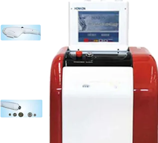
Pmise MF-04
One RF treatment handpiece(Cooling RW-VI with both Mono-polar and Bi-polar functions) and one E-light treatment handpiec…
View details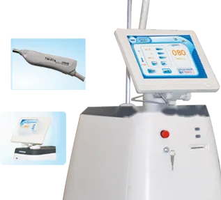
Pmise LN-01
Super compressor cooling system ensures long working time User-friendly operation interface.Direct energy output, less e…
View details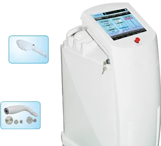
Pmise PE-01
Safe:Single pulse of HPT (Homogeneous Pulse Technology) delivers average energy and avoids the epidermis burns.Efficient…
View details